-
Home


-
Sight Issues
-
Looking After Your Sight
- How Your Eyes Work
How Your Eyes Work
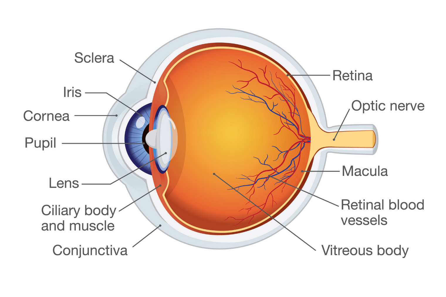
Anatomy of the human eye
The eye is positioned in a socket called the orbit, and is moved by extraocular muscles which are attached to the white part of the eye called the sclera. The sclera provides a tough outer layer that protects the whole eyeball.
The surface of the eye and inner surface of the eyelids are covered with a clear membrane called the conjunctiva, and the eye is lubricated by tears.
When we see something, this is a result of light bouncing off our surroundings and entering our eye.
First of all, the light passes through the dome shaped transparent front layer of the eye called the cornea, which helps to focus the light.
Behind the cornea is a space called the anterior chamber, which is filled with a fluid called aqueous humour, constantly produced by the ciliary body and drained from the eye to maintain a constant eye pressure.
Some of the light that passes through the cornea then enters the eye through an opening called the pupil. The size of the pupil, and the amount of light let in, are controlled by the coloured ring of tissue surrounding the pupil, called the iris.
The light then passes through a transparent tissue called the lens, which fine-tunes the focusing of the light already done by the cornea onto the retina at the back of the eye. The ciliary muscles change the shape of the lens to help the eye focus on objects that are different distances away.
The retina is a layer of tissue that is sensitive to light, which lines the back of your eye. To get to the retina, the light passes through a jelly-like substance called vitreous humour, which fills the space between the lens and the back of the eye, nourishing the eye and helping it to maintain its shape.
Once the light reaches the retina, light sensing nerve cells, called photoreceptors, turn the light into electrical signals. There are two types of photoreceptors: rods, which perceive black and white and allow us to see when light levels are low, and cones, which provide central detailed vision. Cones are found primarily in a tiny, specialized part of the retina called the macula, which allows us to see in detail in the centre of our vision. Rods are found primarily in the peripheral retina, around the macula, which allows us to see with our peripheral (side) vision, but in less detail.
The electric signals created by the photoreceptor cells pass down the optic nerve, which is composed of millions of nerve fibres, to the visual cortex of our brain, where they are converted into the images we see.
Read next
Looking After Your Sight